Click on a thumbnail from above or a bone on the 3D window in order to select.
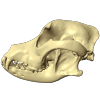
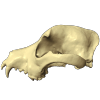
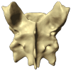
Skull
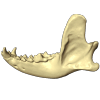
Mandible
×
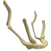
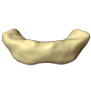
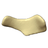
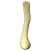
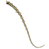
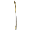
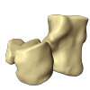
Hyoid
Hyoid
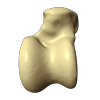
Basihyoid
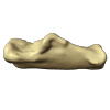
Ceratohyoid
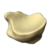
Epihyoid
Stylohyoid
Thyrohyoid
Click on a thumbnail from above or a bone on the 3D window in order to select.
Basihyoid
Ceratohyoid
Epihyoid
Stylohyoid
Thyrohyoid
×
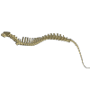
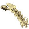
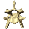
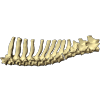
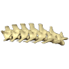
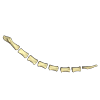
Vertebrae
Vertebrae
Cervical
Atlas (C1)
Axis (C2)
Cervical 6
Thoracic
Lumbar
Sacrum
Caudal
Click on a thumbnail from above or a bone on the 3D window in order to select.
Cervical
Thoracic
Lumbar
Sacrum
Caudal
×
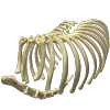
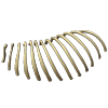
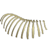
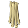
Rib & Sternebrae
Thoracic Cage
Left Rib
Right Rib
Sternebrae
Click on a thumbnail from above or a bone on the 3D window in order to select.
Left Rib
Right Rib
Sternebrae
×
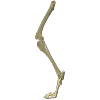
Thoracic Limb
Forelimb
Scapula
Humerus
Radius
Ulna
Radial Carpal
Accessory Carpal
Ulnar Carpal
Distal Carpal
Metacarpal
Proximal Phalanx
Middle Phalanx
Distal Phalanx
Click on a thumbnail from above or a bone on the 3D window in order to select.
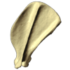
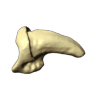
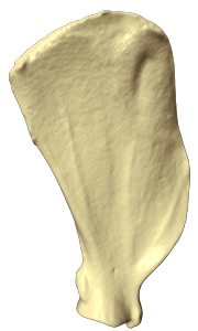
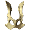
Scapula
Medial View of Scapula
Clincal Aspect
The scapula is a flat triangular bone at the top of the shoulder; more commonly known as the shoulder blade.
It consists of 2 surfaces (medial and lateral), 3 borders (cranial, caudal and dorsal) and 3 angles (craniodorsal, caudodorsal and ventral angle).
The pig and horse do not have an acromion. The acromion protects the suprascapular nerve that lies underneath it, therefore, this nerve is susceptible to physical damage in pigs and horses. Sweeney is a condition in horses in which this nerve is damaged.
Labels & Legends
Acromion: An enlarged distal end of the spine. The deltoid muscle attaches here.
Caudal Border: Relatively straight caudal edge of this bone; closest to the tail.
Cranial Border: Curved cranial edge of this bone; closest to the head.
Dorsal Border: Dorsal edge of this bone with bearing a small scapular cartilage in a live animal; closest to the dorsum (back).
Glenoid Cavity: An articular cavity of the scapula. Articulates with the head of the humerus to form a shoulder joint. This cavity is covered by hyaline (articular) cartilage in a live animal.
Infraglenoid Tubercle: A caudally pointing process found close to the caudal aspect of the glenoid cavity. Articulates with the joint capsule of the shoulder joint.
Infraspinous Fossa: A depressed area caudal to the spine. It serves as a point of attachment for infraspinatus muscle.
Serrated Surface: A rough dorsal portion of medial surface of this bone. The serratus ventralis muscle attaches here.
Spine: A bony ridge that divides the lateral surface of this bone into the supraspinous and infraspinous fossae.
Subscapular Fossa: A smooth portion of the medial surface of this bone. The subscapularis muscle attaches here.
Supraglenoid Tubercle: A cranially pointing process found close to the cranial aspect of the glenoid cavity. This process serves as a point of attachment for the biceps brachii muscle.
Supraspinous Fossa: A depressed area cranial to the spine. It serves as a point of attachment for supraspinatus muscle.
Radiography Positioning
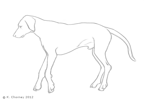
The following illustration shows positioning forelimbs for radiographical imaging.
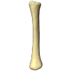
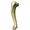
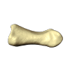
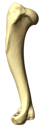
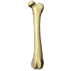
Humerus
Views of Left Humerus: Cranial, Lateral, Caudal, Medial
Some Species Variations
The cat has a small coronoid fossa medial to the radial fossa that accommodates the coronoid process of the ulna during elbow joint flexion.
The cat has a supracondylar foramen near the medial condyle allowing the passage of the median nerve and brachial blood vessels.
There is an intermediate tubercle between the greater and lesser tubercles in the horse's intertubercular groove.
The greater tubercle is subdivided into two parts (cranial and caudal) in ungulates. Ungulates use tips of their digits to bare weight.
Labels & Legends
Deltoid Tuberosity: The tuberosity on the lateral proximal half of the humerus for the attachment of the deltoideus muscle.
Greater Tubercle: Large prominence located craniolateral to the head. This prominence forms a palpable feature known as the point of the shoulder on a live animal. Several shoulder muscles attach here.
Head: The rounded proximal part that articulates with glenoid cavity of the scapula forming the shoulder joint.
Humeral Condyle: The distal extremity of the humerus. Features of the condyle include; capitulum, trochlea, radial and olecranon fossae, and the lateral and medial epicondyles.
Intertubercular Groove: The groove between the greater and lesser tubercle allowing the passage of the tendon of origin of the biceps brachii muscle. In life covered by cartilage to facilitate the smooth movement of the tendon of the biceps brachii muscle.
Lateral Epicondyle: Located on the lateral side of the condyle for the attachment of the lateral collateral ligament and the extensors of the carpus and digits. Functionally called extensor epicondyle.
Lesser Tubercle: Smaller prominence located on the medial aspect of the head.
Line of Triceps Muscle: A raised area between linking the teres minor and deltoid tuberosities. The triceps muscle attaches here.
Medial Epicondyle: Located medial side of the condyle for the attachment of medial collateral ligament and flexors of the carpus and digits. Functionally called flexor epicondyle.
Neck: Constricted portion just below the head.
Olecranon Fossa: Caudal depression that houses the olecranon during the extension of the elbow joint.
Radial Fossa: Cranial depression that accommodates the head of the radius during the flexion of the elbow joint.
Supratrochlear Foramen: The hole between the radial and olecranon fossae found in the dog (occasionally in the pig). It has no known function.
Teres Minor Tuberosity: A proximal lateral tuberosity used for teres minor muscle attachment.
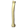
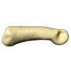
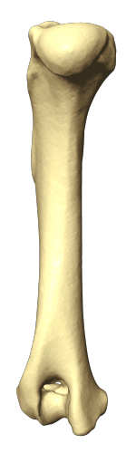
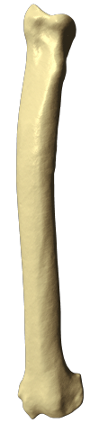
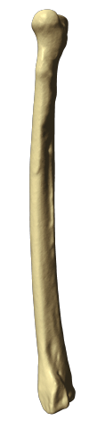
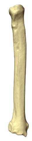
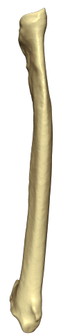
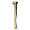
Radius
Views of Left Radius: Cranial, Lateral, Caudal, Medial
Species Variations
The radius and ulna are fused in equidae and ruminants making them inseparable.
The cat has a supracondylar foramen near the medial condyle allowing the passage of the median nerve and brachial blood vessels.
In the horse the styloid process of the ulna completely fuses with the radius. Therefore it is regarded as missing in the horse.
Clinical Aspects of Radius & Ulna
Anconeal and medial coronoid processes may get detached from the ulna in some joint diseases affecting the elbow joint. These can be seen on radiographs of the affected joints.
Labels & Legends
Articular Surface: This is the surface on the head of the radius adapted to articulate with the humeral condyles.
Articular Surface Head: Proximal part of the radius which articulates with the humerus (capitulum) and ulna.
Medial Styloid Process: Pointed end of the distal part of the radius.
Ulnar Notch: This is a small articular facet that accommodates the distal portion of the ulna.
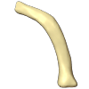
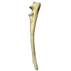
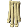
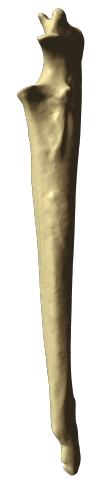
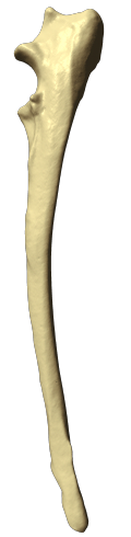
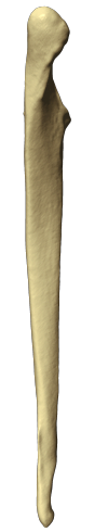
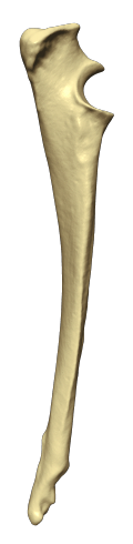
Ulna
Views of Left Ulna: Cranial, Lateral, Caudal, Medial
Species Variations
The radius and ulna are fused in equidae and ruminants making them inseparable.
The cat has a supracondylar foramen near the medial condyle allowing the passage of the median nerve and brachial blood vessels.
In the horse the styloid process of the ulna completely fuses with the radius. Therefore it is regarded as missing in the horse.
Clinical Aspects of Radius & Ulna
Anconeal and medial coronoid processes may get detached from the ulna in some joint diseases affecting the elbow joint. These can be seen on radiographs of the affected joints.
Labels & Legends
Anconeal Process: A large process bordering the proximal margin of the trochlear notch of the ulna. Anconeus muscle attaches here.
Lateral Coronoid Process: Small process on lateral side of the radial notch.
Medial Coronoid Process: Small process on medial side of the radial notch. Medial larger than lateral process.
Olecranon: Caudally pointing tuberosity on the proximal part of the ulna. Serves as an insertion site for major extensors of the elbow joint (e.g Triceps brachii muscle).
Radial Notch: An articular surface for the head of the radius.
Styloid Process: Pointed end of the distal part of the ulna.
Trochlear Notch: A curved articular depression for the humeral trochlea. Bordered my anconeal process proximally and coronoid processes distally.
Radial Carpal
Accessory Carpal
Ulnar Carpal
Distal Carpal I II III IV
Metacarpal I II III IV V
Labels & Legends
Articular Surface:
Proximal Phalanx II III IV V
Middle Phalanx II III IV V
Distal Phalanx I II III IV V
Labels & Legends
Ungual Crest:
Ungual Process:
×
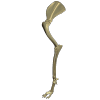
Pelvic Limb
Hindlimb
Os Coxae
Femur
Patella
Tibia
Fibula
Talus
Calcaneus
Central Tarsal
Tarsal
Metatarsal
Proximal Phalanx
Middle Phalanx
Distal Phalanx
Click on a thumbnail from above or a bone on the 3D window in order to select.
The hind limbs have a similar basic pattern to the forelimb. They consist of: femur, tibia and fibula, tarsals, metatarsals, digits or phalanges. The top of the femur moves against (articulates with) the pelvis at the hip joint.
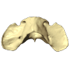
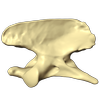
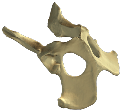
Os Coxae
Dog Os Coxae
Os coxae are made up of four bones; ilium, ischium, pubic and acetabular bones.
Labels & Legends
Acetabular Notch: Notch found on the ventral aspect of the acetabulum.
Acetabulum: A large articulation area with the head of the femur, and divided into Acetabular fossa, Lunatesurface.
Acetabular Fossa: A non-articular depression portion of the acetabulum used for the attachment of the ligament of the head of the femur.
Greater Ischiatic Notch: A notch found on the dorsal border of the body of the ilium. Schiatic nerve and cranial gluteal vessels pass over this notch.
Iliopubic Eminence: Demarcates the ilium from the pubic bone and serves as attatchment site for the prepubic tendon and inguinal ligament.
Ilium: Craniodorsal bone of the os coxae divided into wing and shaft.
Wing: Expanded cranial portion of the ilium.
Shaft (Body): Narrow handle-like part of ilium. Rectus femoris muscle attaches here.
Ischiatic Arch: Caudal margin of the floor of the pelvis formed by the left and right ischiatic bones.
Ischiatic Spine: Found between the greater and lesser schiatic notches and serves for the attachment of the gemellus muscle.
Ischiatic Tuberosity: Laterally projecting process of the ischium used for the attachment of anatomical structures such as sacrotuberous ligament, biceps femoris, semitendinosus and semimemranosus muscles.
Ischium:
Lesser Ischiatic Notch: A notch found on the dorsal border of the ischium. Tendon of internal obturator muscle passes over it.
Obturator Foramen: Largest foramen in the body. Allows the passage of nerves and vessels.
Pelvic Symphysis: A fibrous joint midline on which the two halves of the os coxae are fused ventrally pointing tuber used for the attachment of the prepubic tendon.
Pubic Bone Pectin: Cranially pointing tuber used for the attachment of the prepubic tendon.
Pubis:
Tuber Coxae: Ventral border of the wing (expanded cranial portion of ilium).
Tuber Sacrale: Dorsal border of the wing.
Femur
Labels & Legends
Head: A proximal rounded articular extremity of the femur whose shape is adapted to fit into the acetabulum of the os coxae.
Fovea: A non articular depression found on the head used for the attachment of the ligament of the head of the femur.
Greater Trochanter: It is for the attachment of the middle and deep gluteal muscles as well as the quadriceps femoris muscles.
Intercondylar Fossa: A fossa between the condyles used for the attachments of the cruciate ligaments.
Lateral Condyle: Articular surfaces with the tibia.
Lateral Epicondyle: Muscular and ligamentous attachment site for collateral ligaments, semimembranosus muscle and long digital extensor muscle.
Lateral Supracondylar Tuberosity: Lateral head of the gastrocnemius muscle and the superficial digital flexor muscle attaches here.
Lesser Trochanter: A small process for the attachment of iliopsoas muscle.
Medial Condyle: Articular surfaces with the tibia.
Medial Epicondyle: Muscular and ligamentous attachment site for collateral ligaments, semimembranosus muscle and long digital extensor muscle.
Neck: A constricted portion of the proximal end of the femur distal to the head.
Third Trochanter: Superficial gluteal muscle attaches here.
Trochanteric Fossa: A depression used for the attachment of the internal and external obturator muscles and the gemellus.
Trochlea: An articular surface housing the patella.
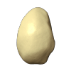
Patella
Tibia
Labels & Legends
Intercondylar Eminence: Process between the two condyles.
Lateral Condyle: Articular surfaces that articulate with the condyles of the femur.
Medial Condyle: Articular surfaces that articulate with the condyles of the femur.
Medial Malleolus: Large process on the distal end of the tibia that serves as a point of attachment for the medial collateral ligament. Palpable in live animals.
Popliteal Notch: Notch between the condyles and allows for the passage of the popliteal muscle tendon.
Tibial Crest: Cranial projection that serves as a point of attachment for the muscles such as biceps femoris, gracillis, Sartorius and semitendinosus.
Tibial Tuberosity: Large cranial prominence of the tibia that serves as a point of attachment for the patellar ligament.
Fibula
Labels & Legends
Head:
Lateral Malleolus:
Talus
Labels & Legends
Trochlea: Articular Surface of Talus.
Calcaneus
Labels & Legends
Sustentaculum: Supports the passage of some flexor tendons.
Tuber: Large tuberosity used for the insertion of the calcaneus tendon. This tendon is made up of tarsal tendons of gracilis, gastrocnemius, and superficial digital flexor muscles.
Central Tarsal
Tarsal I II III IV
Metatarsal I II III IV V
Labels & Legends
Articular Surface:
Proximal Phalanx II III IV V
Middle Phalanx II III IV V
Distal Phalanx II III IV V
×
About
Dog Anatomy project is developed by Veterinary Technician Program and The Sheridan Centre for Academic Excellence in Sheridan College, to provide web-based, interactive digital learning tools for veterinary science students and practitioners.
This project creates scientifically accurate 3D digital models of dog skeletons using a high-definition 3D scanner, and visualizes and simulates 3D digital models to better understand the topographical and functional aspects of dog anatomy.
Under this license, this content is exclusively for non-commercial educational purpose only. It is not permitted users to modify any content in this application, and to publish any content without express written permission of the creators of this work.
Usage
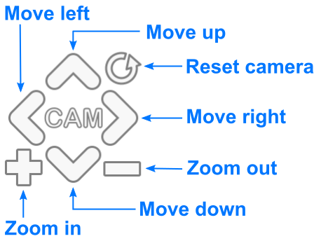
Use the computer keyboard/mouse interface and touch gestures to manipulate the camera.
Mouse Buttons / Wheel : Rotate camera
: Zoom camera in/out
: Move camera
Touches & Keyboards 1-Finger Drag: Rotate camera
2-Finger Drag: Move camera
Pinch: Zoom camera
Arrows: Move camera
+/-: Zoom camera in/out
R: Reset camera